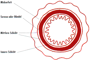
Intestine cross-section
The fine-tissue, microscopic structure of the intestinal wall follows the same basic pattern in all intestinal sections. The innermost layer of the intestinal wall is the intestinal lining, also called the mucosa. The mucosa forms villi or indentations, also called crypts. Intestinal villi are formed in the small intestine, the crypts are typical of the mucous membrane in the large intestine. The mucosa consists of several, very thin layers: a thin layer of mucous membrane muscle, followed by a layer of connective tissue, the submucosa. Very fine blood vessels, lymph vessels and nerve cells end in the submocosa. The next layer is the muscle layer, also known as the muscularis. The muscularis consists of transverse and longitudinal muscle fibers. Because of this muscle arrangement, the intestine can contract both lengthways and crossways. This allows the food pulp to be transported further. The outermost intestinal layer is called the serosa or adventitia and consists of thin connective tissue. In some sections of the intestine, the serosa is formed by the peritoneum, the so-called peritoneum.
The layers of a natural casing can be seen in the cross-section from the outside to the inside. The outermost layer is the so-called gut fat, followed by the serosa, i.e. the epidermis, the submucosa, the muscle layers and finally the mucous membrane with the intestinal villi.
The outer fat is the layer of fat around the intestines, thus also around the intestines.
The serosa is a layer of skin that covers the intestine.
The submucosa is a coarse fibrous, vascular connective tissue sheath and the lower or inner part of the serosa.
The muscle layer is processed into the actual natural intestine. It consists of a longitudinal and transverse fibre layer.
The mucosa is composed of intestinal villi and intestinal glands and is removed during mucus.
